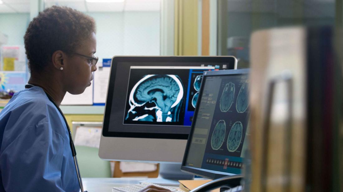
Diagnostic imaging supplies a set of medical techniques and procedures used to visualize the internal structures of the body for the purpose of diagnosing and monitoring various medical conditions. These techniques create detailed images of organs, tissues, bones, and other structures, providing valuable insights for healthcare professionals to make accurate diagnoses and treatment decisions.
Diagnostic imaging encompasses various modalities, each offering unique ways to capture different aspects of the body’s anatomy and function. Some common types of diagnostic imaging include:
- X-rays: X-ray imaging uses low-dose radiation to create images of bones, teeth, and certain soft tissues. It’s commonly used to diagnose fractures, bone deformities, and some lung conditions.
- Computed Tomography (CT or CAT scan): CT scans involve a series of X-ray images taken from different angles, which a computer then assembles into detailed cross-sectional images. CT scans provide more detailed information about various organs, blood vessels, and tissues, making them valuable for diagnosing conditions like tumors, injuries, and vascular issues.
- Ultrasound: Ultrasound imaging employs high-frequency sound waves to produce real-time images of organs and tissues. It’s commonly used to examine developing fetuses during pregnancy, as well as to visualize abdominal organs, the heart, and blood vessels.
- Fluoroscopy: Fluoroscopy is a real-time imaging technique that uses X-rays to observe the movement of internal structures, such as the digestive tract or blood vessels, as they function in real time.
These diagnostic imaging techniques provide valuable information that aids medical professionals in identifying diseases, planning treatments, and monitoring the progress of conditions over time.


